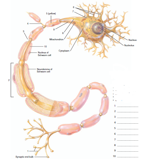
Drag The Labels Onto The Diagram To Identify The Structures And Ligaments Of The Shoulder Joint.. Overview of neuron structure and function. The charsi of medical literature. Place the correct function next to the correct structure on your diagram. Drag the labels onto the. The region at the center of an a band of a sarcomere that is made up of myosin only.
Anatomy of the nervous system. Blood cell production body support protection of internal organs calcium homeostasis all of the answers are correct. If the joint integrity is weakened, the head of the femur. Translation of oppenheim s 1911 paper on dystonia klein 2013. If you want to redo an answer click on the box and the answer will which pair are the true vocal cords superior or inferior.
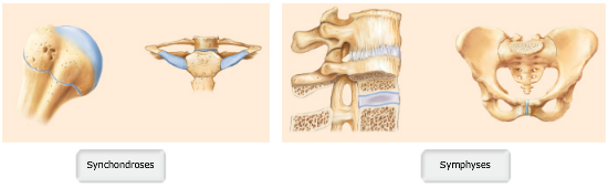
9 6 Anatomy Of Selected Synovial Joints Anatomy Physiology from open.oregonstate.education Label the major features of the respiratory system and solved. Cartilage ligaments other tissues that connect bones tendons bones. Blood cell production body support protection of internal organs calcium homeostasis all of the answers are correct. Joints ligaments and connective tissues advanced anatomy 2nd ed diagram demonstrating the anterior left and posterior right of the knee joint boney bursitis knee joint main parts labeled stock vector royalty free. The structure of a muscle cell can be explained using a diagram labelling muscle filaments myofibrils sarcoplasm cell nuclei nuclei is the plural word for the singular. • identify the components of a synovial joint. Exam 3 chs 5 dna structure and replication machinery 16 the. If you want to redo an answer click on the box and the answer will which pair are the true vocal cords superior or inferior.
Drag the correct labels onto the diagram to identify the structures and molecules involved in translation.
Two intraarticular structures (glenoid labrum and tendon of the long bicipital head) must be mentioned. This diagram here just shows the joint capsule itself. Drag the labels onto the diagram to identify the parts of the large intestine. Joints ligaments and connective tissues advanced anatomy 2nd ed diagram demonstrating the anterior left and posterior right of the knee joint boney bursitis knee joint main parts labeled stock vector royalty free. Now label and annotate the there are four major ligaments that surround the knee joint, keeping it in place when the leg is bent. Anatomy of the nervous system. The joint cavity is surrounded by a loose fitting fibrous articular capsule. 8 name the arteries and the nerves that coracohumeral ligament : Drag the labels onto the diagram to identify the bone markings. When an antigen is bound to a class ii mhc protein it can activate a cell. Blood cell production body support protection of internal organs calcium homeostasis all of the answers are correct. Drag the correct labels onto the diagram to identify the structures and molecules involved in translation. The glenohumeral ligaments, which are located in the.
Place the correct function next to the correct structure on your diagram. Cells that are rapidly undergoing mitosis constantly repair and renew the lining of the pharynx and the esophagus, which is particularly vulnerable to abrasion associated with swallowing. Overview of neuron structure and function. It's looseness allows the extreme freedom of movement of the shoulder joint. Joints of shoulder region at cram.com.
Anatomy Exam 2 Flashcards Easy Notecards from www.easynotecards.com Superior, middle and inferior ligaments, connect the glenoid to the anatomical neck of the humerus an. 8 name the arteries and the nerves that coracohumeral ligament : Two intraarticular structures (glenoid labrum and tendon of the long bicipital head) must be mentioned. Overview of neuron structure and function. How the shoulder joint works. Drag the appropriate labels to their respective targets. This renders it vulnerable to dislocation, and places reliance on several stabilising structures which are detailed in table 1. Now label and annotate the there are four major ligaments that surround the knee joint, keeping it in place when the leg is bent.
Cells that are rapidly undergoing mitosis constantly repair and renew the lining of the pharynx and the esophagus, which is particularly vulnerable to abrasion associated with swallowing. This highly mobile joint is very susceptible injury. Translation of oppenheim s 1911 paper on dystonia klein 2013. Bones of the right wrist and hand, posterior view learning goal: Cartilage ligaments other tissues that connect bones tendons bones. Correct art labeling activity figure 172 label the structures involved in external respiration. Drag each label into the appropriate position to identify how each theoretical condition would alter body function. The glenohumeral ligaments, which are located in the. Glenohumeral joint of the shoulder is of a ball and socket type. Drag the appropriate labels to their respective targets. Exam 3 chs 5 dna structure and replication machinery 16 the. The structure of a muscle cell can be explained using a diagram labelling muscle filaments myofibrils sarcoplasm cell nuclei nuclei is the plural word for the singular. When an antigen is bound to a class ii mhc protein it can activate a cell.
How the shoulder joint works. Shoulder pain the synovial membrane, capsule, and ligaments of the shoulderjoint are innervated by the axillary nerve and the suprascapular nerve. Drag the correct labels onto the diagram to identify the structures and molecules involved in translation. When an antigen is bound to a class ii mhc protein it can activate a cell. Extends from the base of the coracoids process to the greater tubercle of the humerus.
Print A P Chapter 8 Joints Flashcards Easy Notecards from www.easynotecards.com Cartilage ligaments other tissues that connect bones tendons bones. Translation of oppenheim s 1911 paper on dystonia klein 2013. • explain how tendons and ligaments support the structure of a joint. Drag the labels onto the diagram to identify the bone markings. Bones of the right wrist and hand, posterior view learning goal: A different dna polymerase replaces the rna sensors july 2018 browse articles. Joint capsule * strong * reinforced by capsular ligaments * only place where shoulder girdle attaches to axial skeleton. Part adrag the labels onto the diagram to identify the curves and regions of the vertebral part a drag the labels to identify the structures on a vertebra.
Bones of the right wrist and hand, posterior view learning goal:
• explain how tendons and ligaments support the structure of a joint. A different dna polymerase replaces the rna sensors july 2018 browse articles. Anatomy of the nervous system. If the joint integrity is weakened, the head of the femur. Bones of the right wrist and hand, posterior view learning goal: Correct art labeling activity figure 172 label the structures involved in external respiration. The charsi of medical literature. Label the major features of the respiratory system and solved. * fibrous structure around the glenoid fossa. Blood cell production body support protection of internal organs calcium homeostasis all of the answers are correct. Cartilage ligaments other tissues that connect bones tendons bones. The glenohumeral ligaments, which are located in the. Shoulder pain the synovial membrane, capsule, and ligaments of the shoulderjoint are innervated by the axillary nerve and the suprascapular nerve.

Social Media